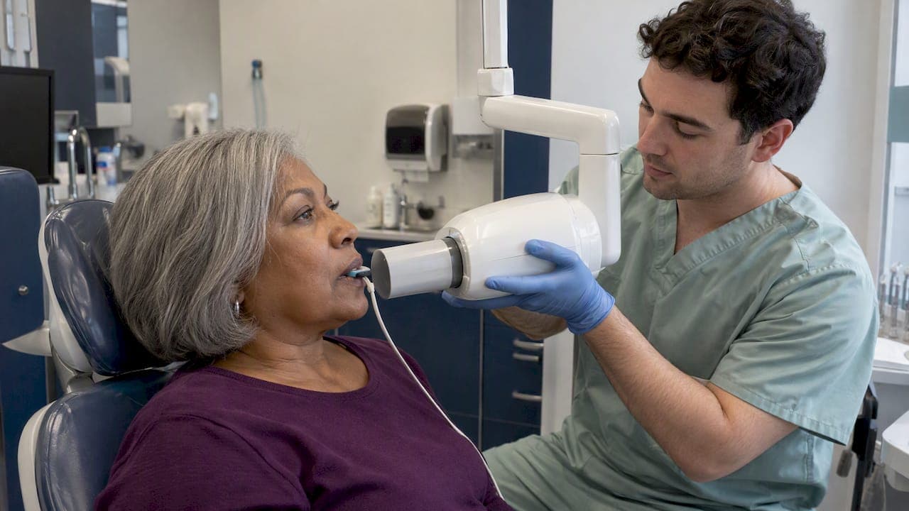
Dental x-rays are safe when a dentist takes them for a reason and takes them well. The dose from a routine set is tiny, and the risk of a missed cavity, infection or bone loss is usually larger than the risk of the image. The real questions are how much radiation you get, how often you need images, and what a careful office does to keep exposure low. This guide answers all three with numbers from the American Dental Association (ADA), the FDA and other primary sources.
TL;DR
- A routine intraoral dental x-ray delivers roughly 1 to 8 microsieverts, typically less than one day of natural background radiation.
- There is no universal schedule. Your dentist sets the interval from your age, cavity history and gum health, anywhere from about 6 months to 3 years between bitewings.
- Pregnancy is not a reason to skip needed dental care. Tell your dentist you are pregnant, and ask whether the image can wait or is needed now.
- Digital sensors, rectangular collimation and good positioning protect you more than a lead apron alone. A careful office also avoids retakes.
- Never demand an x-ray, and never fear a necessary one. Ask your dentist what the image is for.
Table of Contents
- How much radiation is in a dental x-ray?
- What makes a dental x-ray safe in practice
- How often do you really need dental x-rays?
- Dental x-rays during pregnancy
- What good practice looks like at the chair
- Our perspective: how we think about imaging
- When more imaging is worth it
- Questions to ask your dentist
- Exams and x-rays in Washington, DC
- Frequently asked questions
- Sources
How much radiation is in a dental x-ray?
The ADA reports that a routine intraoral x-ray gives an effective dose of about 1 to 8 microsieverts (µSv). That is usually less than one day of the natural background radiation everyone absorbs from soil, air and cosmic rays. Bitewing images in children have been measured as low as 0.77 µSv per image.
A microsievert is hard to picture, so here is the practical reading:
| Image type | What it shows | Typical use |
|---|---|---|
| Bitewing | Crowns of upper and lower back teeth together | Cavities between teeth, bone level |
| Periapical | One or two whole teeth, crown to root tip | Infections, root problems, pain in one spot |
| Panoramic | The whole jaw on one image | Wisdom teeth, implant planning, overall survey |
| CBCT (3D) | A three-dimensional volume | Implants, complex surgery, airway or nerve questions |
The first three are standard 2D images. CBCT uses more radiation than a single intraoral image, which is why it should answer a specific question. We cover that decision in our guide on when a dental CBCT scan is worth it.
Dental imaging in the bigger picture
Dental imaging accounts for less than 1 percent of the collective annual dose from all medical imaging, according to the ADA's journal. That does not make it free of risk. All ionizing radiation carries a small theoretical chance of harm, and there is no technical threshold below which dose counts as zero risk. Safety in dentistry means managing exposure so that every image earns its dose. That idea has a name: ALARA, or As Low As Reasonably Achievable.
What are the side effects?
You will not feel anything, and a routine dental x-ray has no short-term side effects at these doses. The concern is the very small long-term, statistical risk that comes with any ionizing radiation. That is why the question "is this image justified?" matters more than the question "is the machine safe?"
What makes a dental x-ray safe in practice
Safety comes from several layers working together. The machine is only one of them.
Justification: the first and most important step
According to the ADA and the International Atomic Energy Agency (IAEA), the first safeguard is justification. Before any exposure, the dentist asks whether the information is truly needed for diagnosis and whether it can be obtained without radiation. The FDA's guidance on selecting patients for dental radiographs says the same thing in plainer terms: its guidelines are not a substitute for a clinical exam and health history. The dentist should weigh your symptoms, oral and medical history and risk factors first.
Digital sensors
Digital imaging reduces dose by 40 to 60 percent compared with film, according to the IAEA. Most offices now use digital sensors, and the image appears on screen in seconds.
Digital is not automatically risk-free, though. Because a blurry image can be deleted and retaken in seconds, a poorly trained operator can end up giving more total exposure than a careful one. This is the quiet weakness of digital radiography. It does not come from the dose per image. It comes from the habit of retaking. The fix is training and careful positioning so the first image is the right one.
Rectangular collimation
Collimation limits the x-ray beam to the area being imaged. A rectangular collimator shapes the beam to match the sensor instead of exposing a wider round area of your face. Rectangular collimation, together with digital sensors and proper positioning, is now seen as the core of patient protection.
Lead aprons and thyroid collars
This surprises many patients. The ADA now explains that lead aprons and thyroid collars can accidentally block the primary beam, which produces a poor image and a repeat exposure that raises your total dose. Modern practice leans on sensors, collimation and positioning instead. If your dentist does not drape you in lead, that is not a lapse. It is current guidance. Our own explanation, what digital dental x-rays mean for you, walks through the change.
If you prefer an apron, say so. A reasonable office will discuss it with you and explain the tradeoff.
Equipment rules and leakage radiation
The x-ray tube itself is regulated. Rules vary by state, so check your own state's code. As one example, Virginia's code requires that leakage radiation measured one meter from the source not exceed 0.88 milligray in one hour, meaning radiation escaping from the tube housing rather than the beam itself. Similar limits exist in other states. The purpose is that even an aging unit protects patients and staff. Regular inspection and quality control are part of running an office responsibly.
How often do you really need dental x-rays?
There is no one-size-fits-all interval, and you should be wary of any office that says there is. The ADA and the American Academy of Oral and Maxillofacial Radiology published consensus recommendations in 2025 that begin with a thorough evaluation of your history and clinical findings. Frequency follows risk.
As a general reading of that work, recall bitewings fall into two broad groups:
| Your situation | Typical bitewing interval |
|---|---|
| No active cavities and no risk factors | Roughly every 24 to 36 months |
| Active disease or higher risk | Roughly every 6 to 18 months |
Those ranges describe populations, not you. The ADA's own page says how often images are taken depends on your current oral health, age and other specific factors.
Are yearly dental x-rays necessary?
For many low-risk adults, no. A fixed yearly set for everyone is a habit, not a medical rule. For someone with a history of cavities, gum disease, dry mouth or new restorations, annual images may be entirely reasonable. The question to ask is: "What are you looking for, and what would change in my treatment if you find it?"
What changes your interval
- Cavity history. New or recurring decay between teeth is hard to see without bitewings.
- Gum health. Bone loss around teeth shows on x-rays, not in the mirror. Our page on periodontal care and general dentistry explains how we track it.
- Age. Children's teeth change quickly and their tissues are more radiosensitive, which is one reason a published study on pediatric dental radiographs stresses careful dose reference values for them.
- Symptoms. Pain, swelling or a loose tooth can justify an image regardless of your schedule.
- Planned treatment. Implants, root canals, orthodontics and crowns each need baseline images.
- A new office. A new dentist may ask for recent images from your previous office instead of retaking them. Asking is reasonable.
The interval for your cleanings is a separate decision. See our risk-based guide to cleaning intervals for how that works.
Children and sensory needs
Children need images less often when their risk is low, but they should not go without when decay or development questions arise. For kids who struggle with the chair, the sensor or the bite block, preparation helps. Practicing the steps at home with dental social stories for autistic children can make the visit calmer and the first image more likely to be right, which also avoids retakes. Our page on children's oral health describes our approach to young patients.
Dental x-rays during pregnancy
This is one of the most common worries, and the guidance is reassuring. The American College of Obstetricians and Gynecologists (ACOG) has said that dental x-rays and cleanings are safe during pregnancy, and it urged ob-gyns to encourage a dental visit early in prenatal care. Pregnancy also changes gum tissue, so untreated problems can get worse.
What to do in practice:
- Tell the dental team you are pregnant, or might be, before any imaging.
- Ask whether the image is needed now. Routine recall images in a low-risk patient can often wait until after delivery. Images for pain, swelling or an infection should not wait.
- Ask about the technique. Digital sensors, a rectangular collimator and careful positioning keep the dose small and aimed away from the abdomen.
- Talk to your obstetrician if you have a high-risk pregnancy or specific concerns. They know your history, and we are glad to coordinate.
If you have pain or swelling, call a dentist. An untreated infection is a bigger risk than a single small image.
What good practice looks like at the chair
You can tell a lot about an office from how it handles imaging. Look for these signs:
- The dentist explains why an image is needed before taking it.
- Images are tied to your risk, not to a calendar.
- The office uses digital sensors and rectangular collimation.
- Staff are trained in positioning, so retakes are rare.
- Images are reviewed with you on screen, so you see what the dentist sees.
- Prior images are requested and reused where it makes sense.
- You get a clear written treatment plan after the findings.
The IAEA stresses that education and training of dental professionals plays a key role in quality and safety in dental radiology. Equipment is only half of the answer.
Our perspective: how we think about imaging
At DC Implant & Cosmetic Dentistry, we treat an x-ray as a diagnostic decision, not a routine ritual. Dr. Kambez Shukoor is a U.S. Navy veteran, and military medicine taught a habit we still use: you do not gather information you will not act on, and you do not skip information you need.
Because we perform everything from exams to surgical implants in-house, we do not hand you off to a separate imaging center for most questions. That helps us keep the number of images down, since the person planning your treatment is the person who sees the image. It also lets us do thorough imaging and up-front planning when the treatment calls for it, such as implants, where detail matters.
We would rather explain an image than just take it. If you ask why we need one, you should get a straight answer, and if the answer is "we do not, not yet," we will say that.
— Jake
When more imaging is worth it
Some treatments really do need more than bitewings. Implants require knowing bone height, width and the position of nerves and sinuses. Extractions of impacted teeth, bone grafting and complex root canals often need a panoramic or 3D view. In these cases, the extra dose buys information that makes surgery safer, which is the justification principle working as intended.

If you are planning tooth replacement, our page on dental implants, extractions and bone grafting explains how imaging fits into planning. For cancer concerns, imaging is not a screening substitute: an oral cancer screening is a visual and tactile exam.
Questions to ask your dentist
- What are you looking for with this image?
- Would the result change my treatment?
- Can you use images from my previous dentist?
- Do you use digital sensors and rectangular collimation?
- When will I need the next set, and why?
- Is there a reason to take 3D imaging, or will 2D do?
A good dentist welcomes these. If you feel brushed off, that tells you something.
Exams and x-rays in Washington, DC
If you want a dental exam with clear reasoning behind every image, we offer exams and x-rays at our Washington, DC office, with same-day appointments available. You will get a written treatment plan, and everything from checkups to implants is done in-house. See our exams and x-rays page for the types of images we take, or visit our general dentistry page to book. We do not publish prices for these services, so please call for details about cost, insurance and financing.
Frequently asked questions
Is a dental x-ray safe every year?
For patients who need them, yes: the dose is very small, at roughly 1 to 8 µSv per routine image according to the ADA. But many low-risk adults do not need yearly bitewings. Your dentist should base the interval on your risk and explain it.
Are digital dental x-rays safer than film?
Yes, they cut dose by about 40 to 60 percent according to the IAEA. The caveat is that digital makes retakes easy, so training and positioning still matter. A careful office gets the image right the first time.
Do I need a lead apron?
Not necessarily. The ADA notes that aprons and thyroid collars can block the beam and cause repeat images. Sensors, collimation and positioning now do most of the protecting. If you want an apron, ask, and your dentist can discuss it with you.
Can I have dental x-rays while pregnant?
ACOG has said dental x-rays are safe during pregnancy. Routine images can often wait, but images for pain or infection should not. Tell your dentist you are pregnant, and ask what is needed now.
Sources
- X-Rays/Radiographs, American Dental Association
- ADA and AAOMR patient selection for dental radiography and CBCT, JADA
- Dental radiation and collective medical imaging dose, JADA
- The Selection of Patients for Dental Radiographic Examinations, FDA
- Improved justification and optimization of dental imaging, IAEA
- 12VAC5-481-1601, general requirements for diagnostic x-ray systems, Virginia
- Radiation exposure and frequency of dental radiographs in children, PMC
- ACOG: Dental x-rays, prophies OK during pregnancy, DrBicuspid