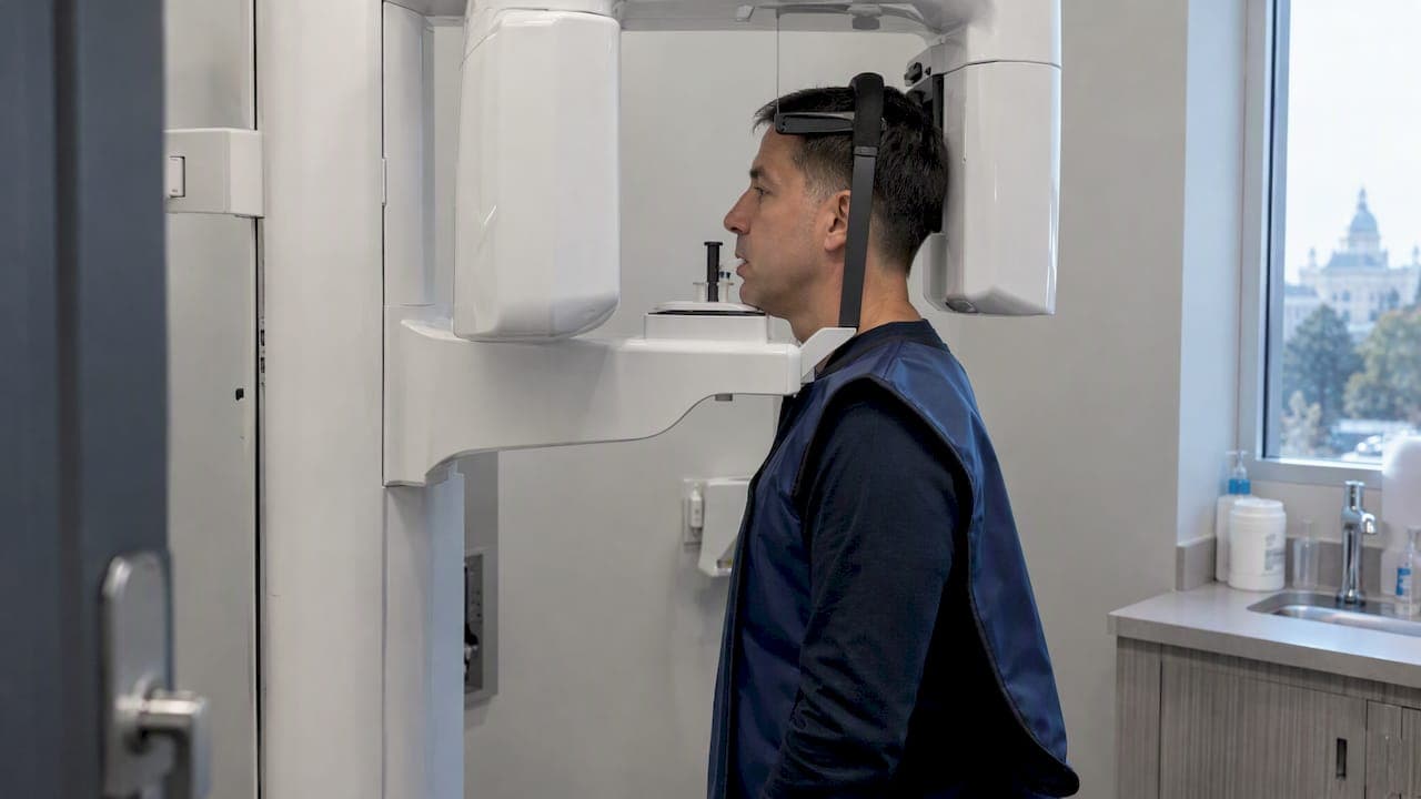
A dental CBCT is a 3D cone-beam X-ray used only when 3D detail changes treatment decisions, for example implant planning or complex root canal problems. It builds a three-dimensional picture of bone and nerve pathways that regular 2D X-rays can't show, which is why dentists reach for it in specific, higher-stakes cases rather than for every checkup.
TL;DR:
- CBCT scans provide detailed 3D images crucial for planning implants, locating impacted teeth, and assessing fractures, but are unnecessary for routine checkups.
- The typical scan takes 10 to 70 seconds and produces a DICOM file; image quality varies with machine settings and field of view.
- Radiation doses depend on the scan size, with small fields offering minimal exposure, especially in children and pregnant patients, following the ALARA principle.
- Cost varies widely based on coverage area and provider, and insurance often covers only specific procedures or parts of the treatment.
- Complex cases benefit from professional interpretation by trained radiologists or specialized dentists to ensure accurate diagnosis and treatment planning.
Table of Contents
- What is dental CBCT and how does it work?
- Common clinical uses of CBCT in dentistry
- What to expect: preparation, the scan visit, and results delivery
- Radiation, safety, and when CBCT is justified
- Cost and insurance: what patients should expect
- Who interprets CBCT images and how you get the report
- Limitations and alternatives to CBCT
- How an advanced dental practice uses CBCT in real cases
- Balancing diagnostic benefit and safety
- How DC Implant & Cosmetic Dentistry can help
- FAQ
- Sources
What is dental CBCT and how does it work?
Cone-beam computed tomography uses a cone-shaped X-ray beam that rotates once around the head, capturing a series of projections and reconstructing them into a 3D volume. According to Radiologyinfo, the detector generates roughly 150 to 200 individual 2D images in a single rotation, and software stitches them into a model you can rotate and slice on screen. A full-mouth scan usually takes between 10 and 70 seconds depending on the area covered, as the FDA notes.
CBCT isn't the same thing as a hospital CT scanner. It delivers sharper detail of bone, teeth, and the jaw than medical CT for this kind of dense tissue work, but it trades away some of medical CT's ability to distinguish soft tissue. Compared with a standard panoramic or periapical X-ray, CBCT adds depth: you see height, width, and thickness instead of one flattened image.
A few practical trade-offs worth knowing:
- Radiation dose shifts with the field of view, so a small scan of one jaw segment delivers far less exposure than a full-arch image.
- Soft-tissue contrast is limited, so CBCT reads bone and teeth well but tells you less about muscles, glands, or ligaments.
- Image quality can be affected by machine settings, meaning two CBCT units from different manufacturers won't always produce identical resolution.
Common clinical uses of CBCT in dentistry
CBCT earns its place in dentistry by answering questions 2D imaging can't. Peer-reviewed dental radiology literature describes CBCT's value for hard-tissue mapping across several recurring scenarios.
- Implant planning: CBCT measures available bone volume, maps nerve position, and checks sinus proximity before a surgeon places an implant.
- Endodontics: it can reveal extra root canals, vertical root fractures, or infection that persists after a root canal looks finished on a flat film.
- Impacted or ectopic teeth: oral surgeons use it to locate wisdom teeth or displaced teeth relative to nerves and adjacent roots before extraction.
- Trauma and jaw fractures: a 3D view clarifies fracture lines that a 2D image might hide behind overlapping bone.
- TMJ evaluation: it assesses the bony structure of the jaw joint, though it has limits for the joint's soft cartilage disc.
- Tumor or pathology localization: it helps pinpoint the extent of a cyst or lesion in the jawbone.
- Orthodontic and orthognathic planning: complex bite or jaw surgery cases sometimes need full skeletal detail that flat films can't provide.
What to expect: preparation, the scan visit, and results delivery
The appointment itself is short and mostly passive. You will likely be asked to remove earrings, necklaces, glasses, and any removable dental appliances, though the FDA notes you should still bring removable appliances with you since your dentist may need to examine them separately.
Positioning depends on the machine. Some CBCT units scan you standing or seated upright in a chair, others use a table similar to a hospital CT. Either way, you'll be asked to stay still and sometimes bite down on a small guide while the scanner rotates around your head.
- The scan itself runs about 20 to 40 seconds for a full volume, per RadiologyInfo.org.
- Clinics affiliated with teaching programs, such as the University of Michigan's CBCT imaging service, report total appointment times of around 20 minutes once positioning and image verification are included.
- Images are often viewable on screen almost immediately, with a formal written report following afterward.
- Expect delivery as a DICOM file you can share with another provider, plus a written radiology report explaining what the scan shows.
Radiation, safety, and when CBCT is justified
CBCT uses more radiation than a routine dental X-ray, but a fraction of what a full medical CT scan delivers. The exact dose depends heavily on the field of view and the specific machine and protocol used.
A peer-reviewed dosimetry meta-analysis found adult effective doses vary widely by protocol: small fields of view often deliver low microsievert doses, while medium and large fields of view may involve higher doses that vary widely with equipment and protocols depending on the equipment and settings used. That range is the reason dentists don't default to the largest scan available.
This is where the ALARA principle comes in: As Low As Reasonably Achievable. The FDA and professional dental organizations recommend clinicians select the smallest field of view and lowest exposure settings that still answer the clinical question, rather than scanning a wider area than necessary out of convenience.
A few groups warrant extra caution:
- Children are more radiosensitive than adults, so pediatric CBCT use should be limited to cases where the diagnostic benefit clearly outweighs the exposure.
- If you're pregnant, tell your dentist before any imaging is scheduled. Many practices defer nonurgent scans until after pregnancy and discuss timing case by case.
- The American Dental Association's guidance in JADA states CBCT should be ordered only when it will meaningfully benefit patient care, with dose and field of view optimized for each case rather than applied as a routine screening tool.
Cost and insurance: what patients should expect
There's no single price for a dental CBCT scan. Cost depends on your provider, your geographic market, and how large an area the scan covers, since a focused single-tooth scan and a full jaw volume aren't priced the same.
Insurance coverage is inconsistent. Many dental plans don't cover CBCT as a routine benefit, and some only reimburse it when it's bundled into a larger procedure claim, such as implant surgery, rather than billed as a stand-alone scan.
- Ask your provider for a written estimate before the scan, not after.
- Confirm whether the imaging center or dental office is in-network with your specific plan.
- Request the procedure code used for billing so your insurer can process preauthorization accurately, as guidance on medical imaging costs recommends confirming coverage details directly with your insurer before the appointment.
Who interprets CBCT images and how you get the report
A CBCT scan produces a large dataset, and reading it correctly takes training beyond what's needed to read a flat 2D X-ray. For straightforward cases, your own dentist may review and interpret the scan. For complex findings, professional guidance suggests a referral to a specialist, such as an oral and maxillofacial radiologist trained specifically in reading these volumes.
- Reports typically include multiple cross-sectional views, measurements of bone or canal structures, and recommended next steps.
- Ask who will personally review your scan and whether that person has specific CBCT interpretation training.
- Ask how long the written report will take and whether you can receive a copy of the DICOM file for your own records or a second opinion.
Limitations and alternatives to CBCT
CBCT is excellent at showing bone and tooth structure, but it isn't the right tool for everything. Its soft-tissue contrast is limited, so a suspected soft-tissue tumor or a TMJ disc problem (the cartilage cushion in the jaw joint, not the bone around it) is usually better evaluated with an MRI or medical CT instead, as dental radiology reviews note.
- Metal crowns, implants, or fillings can create artifacts that distort the image around them, sometimes obscuring the exact detail a dentist is trying to see.
- A routine cavity check or a standard cleaning visit doesn't need 3D imaging. A standard digital X-ray usually answers the question just as well with far less exposure.
- CBCT is reserved for cases where flat imaging genuinely falls short, not as an upgrade for every visit.
How an advanced dental practice uses CBCT in real cases
A practice built around in-house imaging can move from diagnosis to treatment plan in a single visit. At DC Implant & Cosmetic Dentistry, CBCT scans pair with intraoral scanning to plan dental implants and complex oral surgery cases without sending patients elsewhere for imaging. Some practices with same-day appointment availability mean a patient needing implant planning can often get scanned and reviewed in one trip. Ask any practice performing CBCT who will interpret your scan and how quickly you'll get a written report.

Balancing diagnostic benefit and safety
CBCT earns its place when 3D detail genuinely changes a treatment decision, not as a routine add-on. Before any scan, ask your dentist three things: why this scan is necessary, what the dose will be for your specific field of view, and who will interpret the images. Those three questions tell you whether the scan is justified.
— Jake
How DC Implant & Cosmetic Dentistry can help
Comprehensive dental care works best when imaging, planning, and treatment happen under one roof instead of bouncing between offices. DC Implant & Cosmetic Dentistry performs exams, X-rays, and implant planning in-house, so a CBCT finding leads directly into a written treatment plan rather than another referral.

If you're weighing a dental implant, an extraction, or a complex restoration, schedule a consultation for exams and X-rays to see how imaging fits your specific case.
This article is general information, not a substitute for advice from a qualified doctor. Consult a qualified healthcare professional about your own circumstances before acting on anything here.

FAQ
Does insurance cover a CBCT scan?
Coverage varies by plan, and many dental insurers don't cover CBCT as a stand-alone, routine benefit. Confirm with your insurer whether the scan will be covered independently or only as part of a larger procedure, and ask your provider for the billing code used for preauthorization.
Why would a dentist order a CBCT scan?
A dentist orders CBCT when a treatment decision depends on 3D detail that a flat X-ray can't provide, such as mapping bone before an implant or finding a hidden root canal issue. Professional guidance recommends it only when the diagnostic benefit outweighs the added radiation exposure.
What is the cost of a CBCT scan?
There is no single published price for a dental CBCT scan, since cost depends on the provider, the region, and how much of the jaw the scan covers. Ask your dental office for a written estimate and confirm with your insurer whether preauthorization is required before the appointment.
How much does a dental CBCT scan cost?
Pricing isn't standardized and shifts based on field of view, equipment, and local market rates, so the only reliable number is the one your own provider quotes you. Request an itemized estimate and the procedure code in advance so your insurer can confirm what, if anything, it will cover.
Sources
- Dental Cone-beam Computed Tomography | FDA
- Dental Cone Beam CT
- Cone Beam Computed Tomography - Know its Secrets - PMC
- CBCT Imaging Service | University of Michigan School of Dentistry