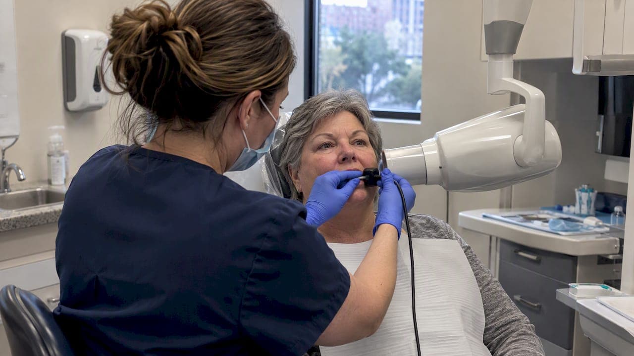
Digital dental X-rays use electronic sensors instead of film to capture images that appear on a screen within seconds. They expose patients to less radiation than older film systems and support the ALARA principle (as low as reasonably achievable) that guides modern dental imaging. Current ADA guidance no longer recommends routine lead aprons, since beam restriction and digital equipment already limit exposure effectively.
TL;DR:
- Typical doses are 1 to 8 microsieverts for intraoral images, 4 to 30 for panoramic scans, while larger CBCT scans can reach 100 or more.
- CBCT should be reserved for cases where two dimensional images lack needed detail, such as implant planning or complex root canal assessment.
- ADA guidance no longer recommends routine lead aprons or thyroid collars; beam restriction limits exposure, while an apron can block the beam and cause retakes.
- Digital sensors can hide overexposure because images may still look acceptable, so ask whether the office adjusts settings for patient size and tracks retakes.
- X ray frequency depends on clinical findings, age, and cavity or gum disease risk, not a fixed schedule; ask whether recent images can be reused.
Table of Contents
- What a digital dental X-ray is and how it works
- Types of digital receptors and typical dental X-ray exams
- Benefits of digital X-rays vs. film for patients
- Radiation exposure and safety: numbers, recommendations, and what patients should ask
- What to expect during X-rays and how often you'll need them
- How digital X-rays help diagnose cavities, bone loss, and implant needs
- Our approach to imaging and patient safety
- How to get digital X-rays or an exam at DC Implant & Cosmetic Dentistry
- FAQ
- Sources
What a digital dental X-ray is and how it works
A digital dental X-ray replaces traditional film with an electronic sensor or plate that converts X-ray energy into a digital image. Instead of waiting for film to be developed in a darkroom, your dentist sees the image on a monitor within a few seconds of exposure, which allows an immediate check for clarity before you leave the chair.
Most routine visits involve small intraoral images taken with a sensor placed inside your mouth:
- Bitewing X-rays show the crowns of upper and lower teeth together, mainly to catch cavities between teeth.
- Periapical X-rays capture an entire tooth, from crown to root tip, to check for infection or bone changes.
- Panoramic X-rays rotate around your head to produce one wide image showing all your teeth, jaws, and sinuses.
- Cone beam CT (CBCT) scans create a three-dimensional image and involve a higher dose, reserved for cases like implant planning or complex root canals.
Types of digital receptors and typical dental X-ray exams
Digital sensors fall into a few categories, and the one your dentist uses affects what the appointment feels like. CCD and CMOS sensors are wired directly to a computer and display images almost instantly, which makes them the fastest option for routine checkups. They tend to be rigid and slightly bulkier than film, so your dentist may use a positioning holder to keep the sensor steady and comfortable against your teeth.
Photostimulable phosphor (PSP) plates work differently. They look and feel more like traditional film, which some patients find more comfortable, but they need to be scanned by a separate reader after exposure rather than showing up instantly on screen.
- Direct sensors (CCD/CMOS) suit quick bitewing and periapical checks where speed matters.
- PSP plates offer a thinner, more flexible option for patients who find rigid sensors uncomfortable.
- Panoramic units handle a full jaw overview in one pass, useful for orthodontic planning or spotting impacted teeth.
- CBCT is recommended only when two-dimensional images don't give enough detail, such as before placing an implant or assessing complex anatomy. A dedicated breakdown of when a CBCT scan is worth it covers the questions worth asking before one is ordered.
Benefits of digital X-rays vs. film for patients
Digital imaging's biggest practical advantage is dose reduction. The FDA notes that digital imaging can lower patient radiation dose by a substantial margin compared with older film systems, depending on the receptor and technique used. That's on top of earlier gains from faster film speeds, since the FDA's film speed data shows switching from D-speed to E-speed film historically cut exposure by a noticeable amount, with F-speed film reducing it further still.
The FDA's analysis shows digital imaging can cut patient dose by roughly 40 to 60% versus older film when the equipment and technique are optimized, a meaningful reduction for anyone who needs imaging regularly.
Beyond dose, digital systems let your dentist enlarge, brighten, or adjust contrast on an image instantly, which helps catch small cavities or bone changes that might be missed on film. Images also transfer electronically between offices, and there's no chemical processing or film waste involved.
- Faster results mean fewer retakes caused by processing errors.
- Easier sharing supports better continuity of care between specialists.
- No darkroom chemicals mean less environmental waste from the practice.
The flexibility cuts both ways, though. Digital sensors have a wide exposure latitude, which means an overexposed image can still look acceptable on screen. Without careful technique, that can lead to what radiation physicists call exposure creep, where dose gradually increases without anyone noticing.
Radiation exposure and safety: numbers, recommendations, and what patients should ask
Dental X-ray doses are small relative to many other imaging exams. According to the IAEA's review of dental radiology, typical effective doses run about 1 to 8 microsieverts for intraoral images, 4 to 30 microsieverts for a panoramic scan, and a wider range for CBCT, from well under 50 microsieverts for a small field of view up to around 100 microsieverts or more for larger volumes.
| Exam type | Typical effective dose |
|---|---|
| Intraoral (bitewing/periapical) | About 1 to 8 microsieverts |
| Panoramic | About 4 to 30 microsieverts |
| CBCT (small to large volume) | Roughly under 50 to 100+ microsieverts |
CBCT remains a major contributor to dental diagnostic radiation, according to the IAEA, which is why it's reserved for cases where a flat 2-D image won't give your dentist what's needed.
The ALARA principle drives most of the dose-lowering steps your dentist should already be using: rectangular collimation to narrow the beam to the size of the sensor, the fastest receptor speed available, and posted technique charts that set exposure parameters by patient size so a child isn't exposed at an adult setting. The ADA's updated guidance found that lead aprons and thyroid collars are no longer routinely necessary, since modern beam restriction already protects tissue outside the exam area, and aprons can sometimes block part of the beam and cause a retake.

An IAEA technical report on digital radiography makes an important point: digital detectors can reduce dose, but if technique and quality assurance are not carefully maintained, doses can increase over time. Operator factors, like how often retakes happen or how carefully a sensor is positioned, matter as much as the technology itself.
Pro Tip: Ask your dental office whether they post technique charts for their X-ray units and how often they need to retake images, since both answers tell you a lot about exposure control.
What to expect during X-rays and how often you'll need them
Getting a digital dental X-ray is quick and involves a predictable sequence.
- You'll be seated in the dental chair, often with a protective drape or no apron at all under current guidance.
- A sensor or plate is positioned inside your mouth, sometimes with a holder to keep it steady and reduce retakes.
- The technician or dentist steps back and takes the exposure, which lasts a fraction of a second.
- The image appears on a monitor within seconds, ready for review.
How often you need X-rays depends on your clinical exam, age, and risk factors like a history of cavities or gum disease, following ADA and AAOMR selection criteria rather than a fixed schedule. You can ask your dentist why a particular image is being recommended, request that prior images be transferred from a previous provider, or decline an X-ray, though declining may limit your dentist's ability to diagnose issues that aren't visible during a visual exam.
How digital X-rays help diagnose cavities, bone loss, and implant needs
Digital X-rays let your dentist see problems that aren't visible to the eye. Cavities forming between teeth often hide from a visual exam but show up clearly on a bitewing image as a dark spot in the enamel or dentin, often before the decay causes any pain.
Bone loss from gum disease is another area where digital imaging matters. Periapical and panoramic images show the bone level around each tooth root, so your dentist can track whether that level is stable or receding over time, a key factor in deciding whether a tooth can be saved.
For implant planning, imaging needs are more detailed. A CBCT scan shows the width, height, and density of the jawbone in three dimensions, along with the location of nerves and sinuses, information a flat 2-D image can't provide. That level of detail helps determine implant size and placement before any surgery happens, reducing surprises once treatment begins.
Digital images also support broader treatment planning when paired with other tools. An intraoral scanner captures a 3-D model of your teeth and bite, which combined with X-ray data gives a fuller picture for anything from a single crown to a full reconstruction.
Our approach to imaging and patient safety
We use CBCT and digital intraoral imaging to support precise implant planning and everyday diagnosis, keeping technique charts and dose optimization at the center of how we take images.
— Jake
How to get digital X-rays or an exam at DC Implant & Cosmetic Dentistry
We offer Exams & X-Rays as part of routine visits, with CBCT scans available when a case calls for implant planning or complex diagnosis rather than as a default step.

If you've had recent X-rays taken elsewhere, bring them or have your previous office send them over. That often means fewer repeat exposures and a faster start to your visit.
- Appointments are available for exams and X-rays.
- Prior images can be reviewed alongside new ones to track changes over time.
- Financing options are available for larger treatment plans.
For background on modern safety guidance and the questions worth asking before any imaging, this patient-friendly overview walks through why lead aprons are no longer routine. Schedule an exam with us to get a clear, written plan before anything else happens.
This article is general information, not a substitute for advice from a qualified doctor. Consult a qualified healthcare professional about your own circumstances before acting on anything here.
FAQ
What are X-rays used for in dentistry?
Dental X-rays help your dentist find problems not visible during a visual exam, including cavities between teeth, bone loss from gum disease, infections at the root tip, and the position of teeth and nerves before procedures like implants or extractions. They're also used to plan orthodontic treatment and check the fit of restorations.
How much does a 3D dental X-ray cost?
Pricing for CBCT scans varies by practice and isn't published as a flat rate, so the most reliable way to get a figure is to ask your dental office directly. At our practice, exam and imaging costs are discussed as part of a written treatment plan before any scan is taken.
Can I refuse dental X-rays?
You can decline dental X-rays, and your dentist should respect that choice while explaining how it may limit their ability to diagnose issues like hidden cavities or bone loss. Ask what a specific X-ray is meant to find and whether a recent prior image could be used instead.
What is a digital scanner in dentistry?
A digital or intraoral scanner captures a 3-D model of your teeth and bite without the putty impressions traditionally used for crowns, aligners, or dentures. It's a separate tool from X-ray imaging, though both are often used together for complete treatment planning.
Are dental X-rays safe during pregnancy?
Dental X-rays are generally considered safe during pregnancy when the exam is justified and standard protective steps are followed, though guidance continues to be refined as techniques improve. A pregnancy-focused guide aligned with current ADA updates covers the specific questions worth raising with your dentist.
Sources
- ADA releases updated recommendations to enhance radiography safety in dentistry
- Dental Radiographic Examinations: Recommendations for Patient Selection and Limiting Radiation Exposure (FDA)
- Radiation protection of patients in dental radiology (IAEA)
- IAEA technical report on digital radiography and patient dose